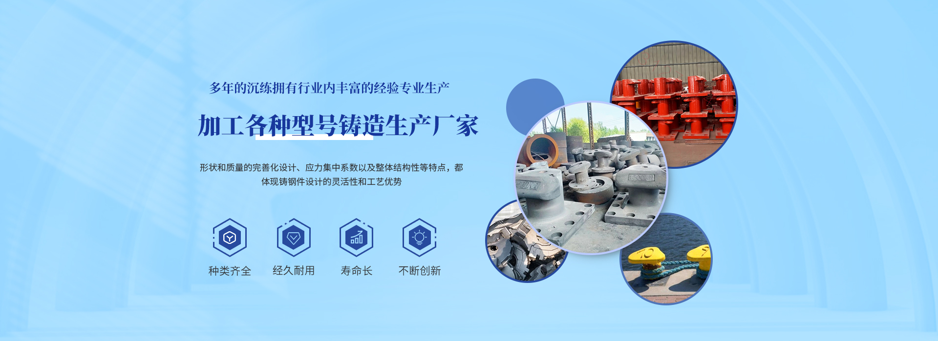
歡迎訪問(wèn)沾化欣瑞康生物科技有限公司網(wǎng)站~
<strike id="eoicc"></strike>
歡迎訪問(wèn)沾化欣瑞康生物科技有限公司網(wǎng)站~

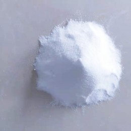
肌酸酐
肌酸酐介紹:別名名稱(chēng):2-Amino-1-methyl-2-imidazolin-4-one 2-Imino-1-methylimidazolidin-4-one 2-Imino-N-methylhydantoin,生化研究。用于鑒定血液中的肌酸酐。本品應(yīng)密封于干燥處保存。 合成方法...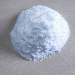
磷酸肌酸二鈉鹽
磷酸肌酸二鈉鹽的用途,主要用途是生產(chǎn)注射針劑磷酸肌酸鈉的重要原料。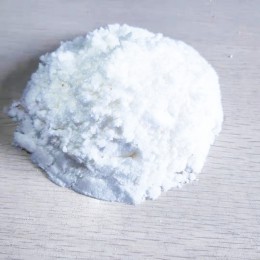
肌酐磷酰氯
本產(chǎn)品是一類(lèi)重要的化學(xué)中間體,具有非常廣泛的用途,在殺蟲(chóng)劑、延緩劑、潤(rùn)滑劑等中均有用到,同時(shí)能合成各種生物活性化合物,接下來(lái)就分析一下七應(yīng)用知識(shí)。 1、合成前列腺累化合物,肌酐磷酰氯在紅細(xì)胞膜不同組織中表現(xiàn)一定的生物活性。 2、利用肌酐磷酰氯合成氨基磷酸酯類(lèi)化合物,并且具有非線...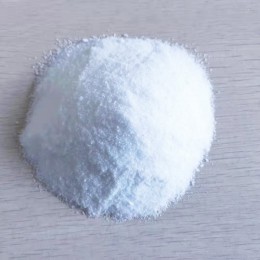
肌酸
肌酸是一種含氮的有機(jī)酸,化學(xué)式為C4H9N3O2,自然存在于脊椎動(dòng)物體內(nèi),能夠輔助為肌肉和神經(jīng)細(xì)胞提供能量。米歇爾·歐仁·謝弗勒爾(Michel Eugène Chevreul)于1832年初次在骨骼肌中發(fā)現(xiàn)肌酸,而后,根據(jù)希臘語(yǔ)“Kreas”(肉),命名為“Creatine”。 肌...

欣瑞康生物科技
公司秉承“團(tuán)結(jié)、協(xié)作、誠(chéng)實(shí)、守信”的經(jīng)營(yíng)理念,堅(jiān)持“以市場(chǎng)為導(dǎo)向,以人才為根本、以創(chuàng)新為動(dòng)力,以品質(zhì)為基礎(chǔ)”的經(jīng)營(yíng)方針,倡導(dǎo)和推行現(xiàn)代化企業(yè)管理模式,以簡(jiǎn)單管理為宗旨,致力于產(chǎn)品研發(fā)及創(chuàng)新,為顧客創(chuàng)造贏利空間,歡迎廣“大客戶(hù)與我們聯(lián)系,成為我們事業(yè)的合作伙伴!
為什么選擇我們
公司秉承“團(tuán)結(jié)、協(xié)作、誠(chéng)實(shí)、守信”的經(jīng)營(yíng)理念,堅(jiān)持“以市場(chǎng)為導(dǎo)向,以人才為根本、以創(chuàng)新為動(dòng)力,以品質(zhì)為基礎(chǔ)”的經(jīng)營(yíng)方針。Professional team
reasonable price
Production equipment
after-sales service
行業(yè)經(jīng)驗(yàn)豐富
欣瑞康生物科技一家集生產(chǎn)、研發(fā)、銷(xiāo)售磷酸肌酸二鈉鹽、肌酐磷酰氯、肌酸酐、磷酸肌酸酐二鈉鹽、磷酸肌酸二納鹽、一 水肌酸等產(chǎn)品的高科技化工企業(yè)。以為客戶(hù)創(chuàng)造持久價(jià)值,為員工創(chuàng)造發(fā)展機(jī)會(huì),為社會(huì)提供環(huán)保的產(chǎn)品為企業(yè)使命。


沾化欣瑞康生物科技有限公司主營(yíng),磷酸肌酸二鈉鹽,肌酐磷酰氯等產(chǎn)品,現(xiàn)貨充足,質(zhì)量好,高純?cè)喜少?gòu),歡迎咨詢(xún)
網(wǎng)站地圖